Composite optical microscope
The study of histology depends on the use of microscopy. Using a light microscope, the histological preparations already stained can be analyzed, so that the histology student must know the basic principles of microscopy. Therefore, a more detailed description of an optical microscope (used in our studies) and the study of some concepts related to optical microscopy become necessary. Finally, the description of other types of microscopes, in addition to the optical microscope, will also be covered.
A compound light microscope (optical) can be simple or composite, the simple microscope having a single lens and only providing a moderately enlarged image of the object being studied, and the composite microscope consists of a set of lenses capable of providing a much bigger increase. The specimen to be observed will be analyzed thanks to the lighting that will pass through it.
The optical microscope is composed of mechanical and optical parts. The mechanical part is the 'support' of the microscope, and consists of a base, whose function is to stabilize the microscope, a column or cannon that extends from the base upwards, supporting the lenses, and a platinum, on which the object to be examined.
The optical parts of interest are attached to the column, above and below the stage, being composed of the eyepiece lenses (one or two eyepieces may be present) and objectives, condenser and mirror. In many microscopes, the mirror and the lamp are safely housed in the base of the instrument.
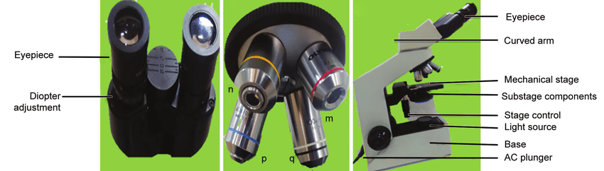
The eyepiece consists of a combination of lenses that are embedded in the upper end of the microscope tube. The recorded value such as 12.5x indicates the magnification of the eyepiece. The objectives (there may be three, four or five) are a combination of lenses attached to the lower end of the microscope tube.
The recorded value, such as 10x, indicates the magnification of the lens. A 10x lens used in combination with a 12.5x eyepiece gives a total magnification of 125x. The different objectives attach to the revolver, which in turn is attached to the lower end of the microscope tube. One objective is exchanged for another by rotating the revolver, so that when one objective replaces the previous one.
The condenser is a combination of lenses located below the stage, whose function is to project a cone of light on the object being observed. The condenser can be raised or lowered by a rack mechanism, so that the light can be focused more or less intensely on the object.
The passage of marginal rays in the condenser is prevented by the iris diaphragm. The diaphragm also regulates the amount of light that leaves the condenser and reaches the object, and its opening can be increased or reduced through manual control.
The mirror below the condenser reflects the light rays emanating from the light source. Located between the mirror and the condenser there is a mobile filter holder.
�
